
Credit: Dr. Hailin Fu Group, Westlake University
Polar-SIM's exclusive polarization helps to resolve structural cracks in supramolecular materials
Polarized structured illumination super-resolution microscopy imaging system (Polar-SIM) is independently developed by Airy Technologies. Based on the structured illumination microscopy (SIM), the system innovatively introduces polarization optical modulation technology, which successfully breaks through the resolution limit of traditional SIM and achieves an ultra-high resolution of about 60 nm. Polar-SIM combines low phototoxicity with high compatibility and can be seamlessly integrated into a wide range of high-end microscopy platforms, providing a state-of-the-art solution for live-cell super-resolution imaging.

More than 20 imaging modes, suitable for cell or tissue imaging at different depths to meet a variety of experimental needs
Faster imaging speed, larger imaging field-of-view, multi-point scanning, ultra-low phototoxicity, long-term tracking, etc

A variety of reconstruction algorithms restore the real structure of the sample to assist scientific new discoveries
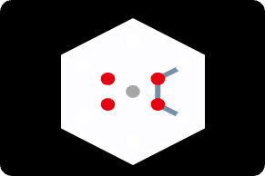
Polar-SIM provides polarization information embedded in fluorescence, helping to reveal the tissue arrangement of cellular structures in specific directions
Polar-SIM provides more than 20 imaging modes (including illustrated modes), compatible to polarized super-resolution imaging, which fully meets the imaging needs of different levels of applications ranging from cells to tissues.
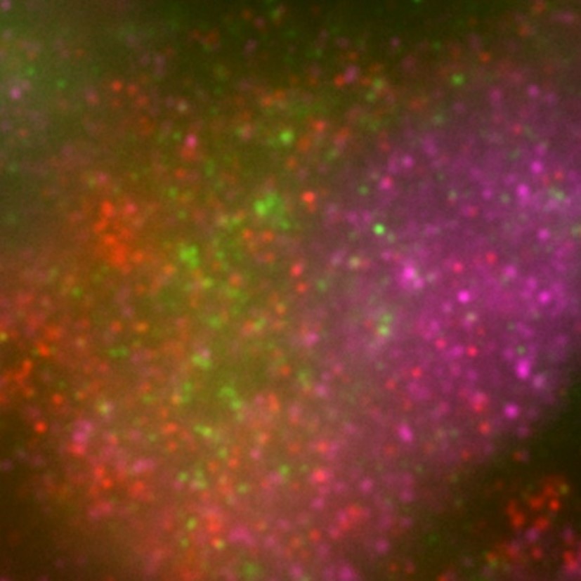
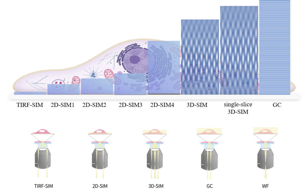
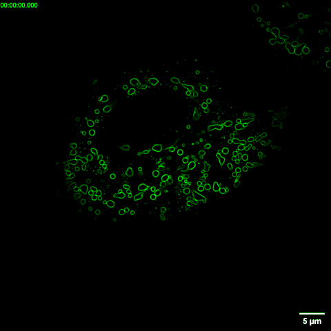
The imaging thickness is ~100 nm near the slide, with excellent signal-to-noise ratio for observing cell membranes at near surface biological structure/phenomena such as cell membrane proteins.
Imaging conditions: 100 x / 1.49, TIRF-SIM mode
488/561/640laser excitation
Sample information: Membrane protein (red/green/magenta)
Sample source: Prof. Weidong Zhao Group, China Medical University
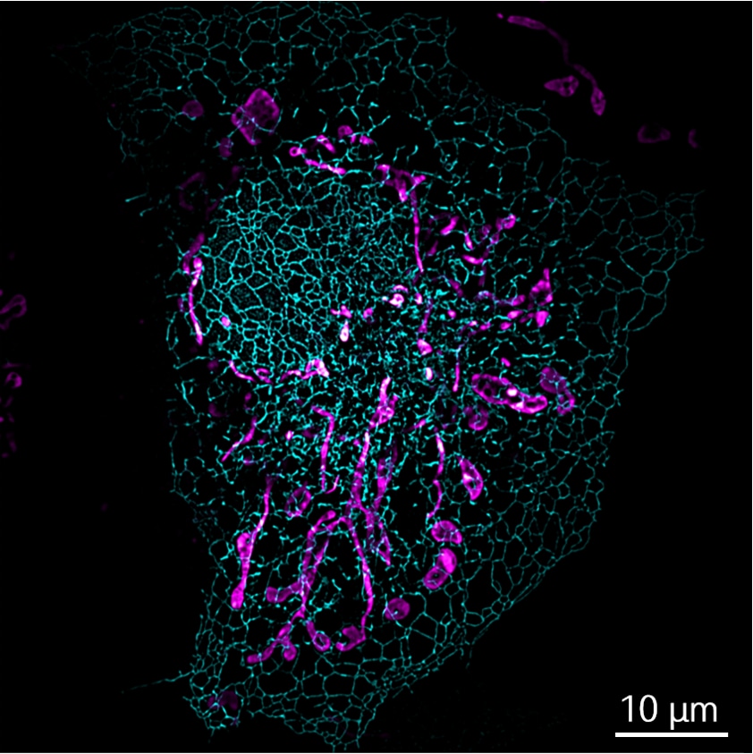
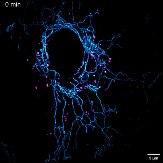
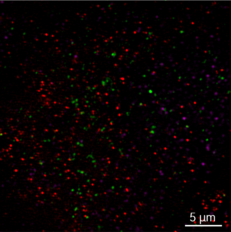
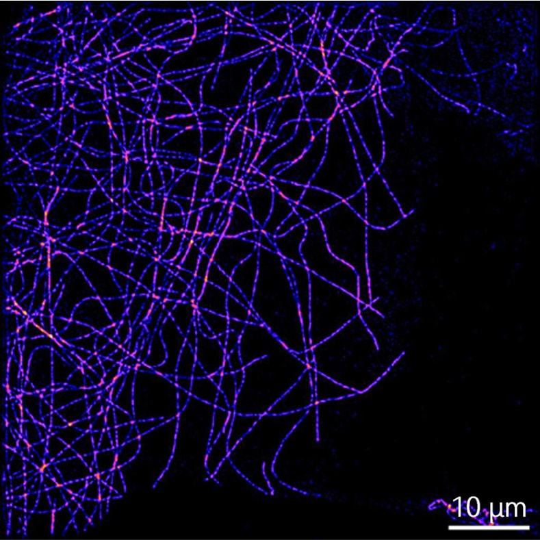
Different levels can be selected according to the thickness of the imaging sample. It is suitable for observing the dynamic processes of organelles in the cytoplasm such as endoplasmic reticulum and mitochondria.
Imaging conditions: 100 x / 1.49, 2D-SIM mode
561/640 laser excitation
Sample information: mitochondria (magenta); microtubes (blue)
Sample source: AiryTech
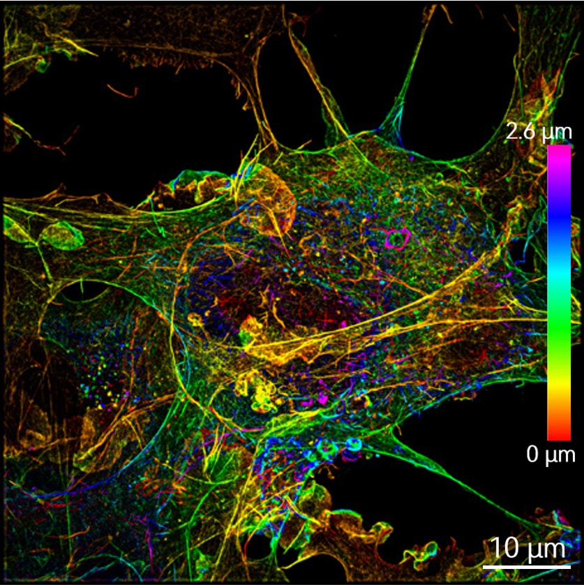
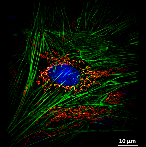
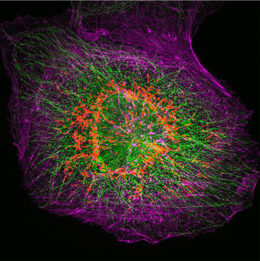
It can achieve double resolution improvement in both lateral and axial dimensions simultaneously. It is suitable for the observation of 3D super-resolution stereoscopic sub-cellular organelle structures, such as centrosomes.
Imaging conditions: 100 x / 1.49, 3D-SIM mode
561laser excitation
Sample information: actin filaments
Sample source: AiryTech
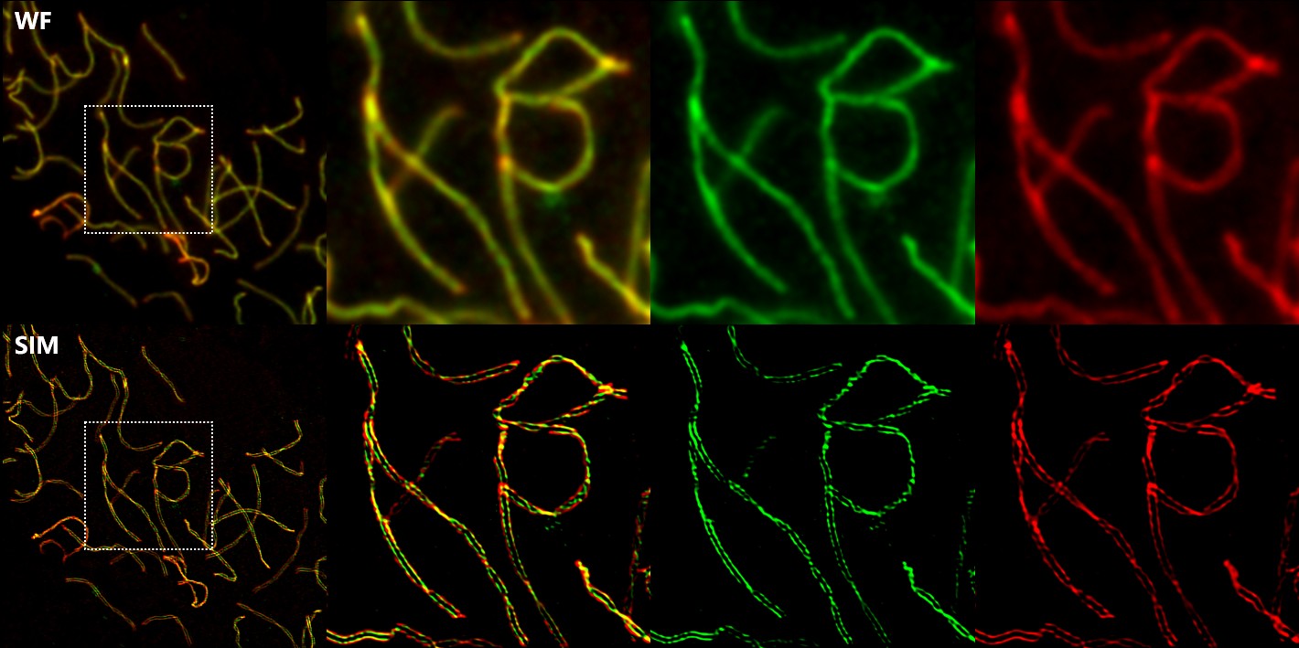
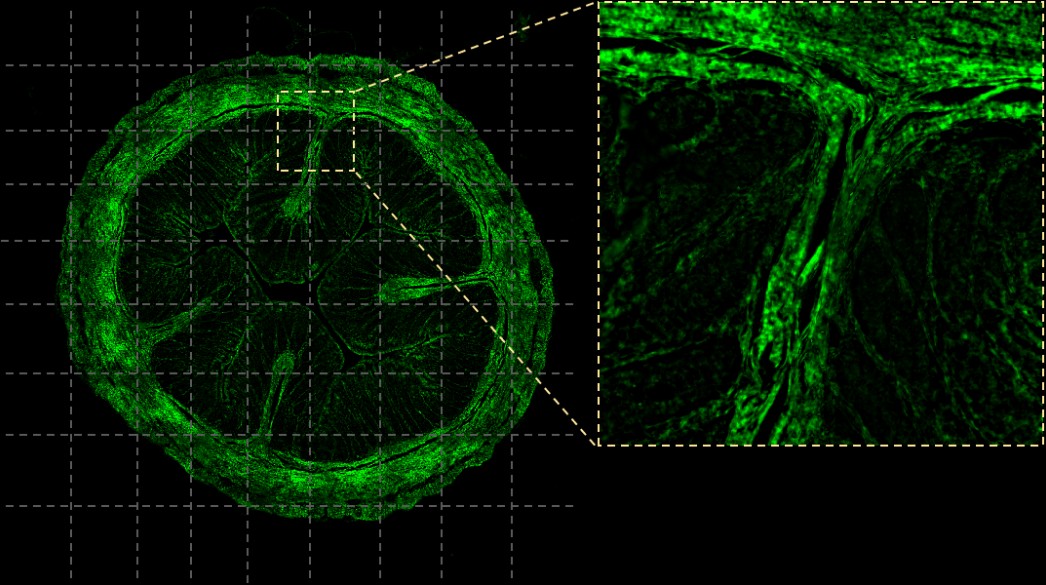
It can effectively remove the out-of-focus background and has the ability to obtain optical sectioning imaging. It is suitable for the observation of thicker biological sample morphology, such as nuclei, tissue sections, etc.
Imaging conditions: 100 x / 1.49, Slingle-slice 3D-SIM mode
561laser excitation
Sample information: microtubules
Sample source: AiryTech
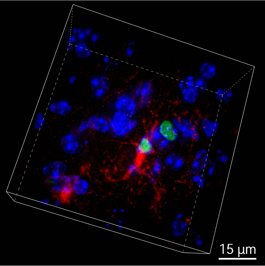
Structured illumination technology not only enables super-resolution imaging, but also has confocal-like optical sectioning capabilities. AiryTech's exclusive structured illumination confocal mode achieves resolution of up to 150 nm and imaging depth of more than 50 μm in just two raw images, meeting the rapid imaging needs of deep tissues. It is suitable for observing small model animals such as biological tissue sections, fruit flies, zebrafish, and nematodes.
Imaging conditions: 100 x / 1.49, GC mode
405/488/561 laser excitation
Sample information: nucleus (blue); Nucleolin protein (green);Filamentous structure (red)
Sample source: Prof. Weidong Zhao Group, China Medical University
With a large imaging field-of-view of 130 x 140 μm (100X), Polar-SIM can cover a wider sample area in a single exposure, significantly improving observation efficiency and data integrity, and avoiding image stitching errors. In addition, the ultra-large imaging field-of-view can simultaneously capture more biological structures or cell dynamics, improving data reliability.
Polar-SIM provides automated multi-point time-lapse imaging that can loop through preset points-of-interest, significantly reducing experimental duplication and greatly increasing the probability of capturing rare events.
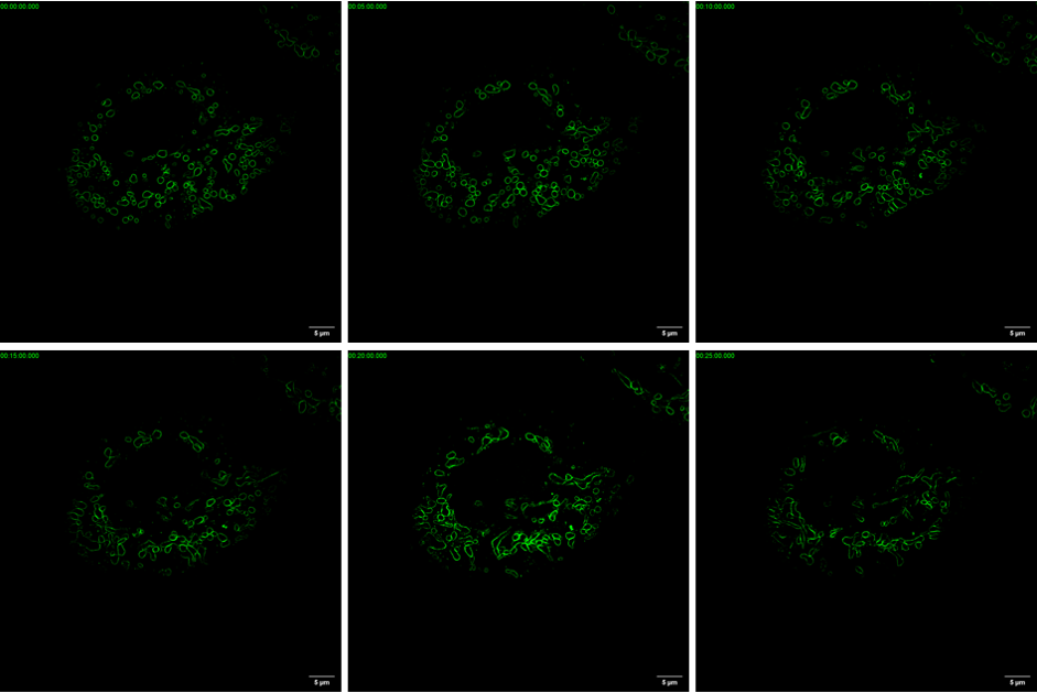
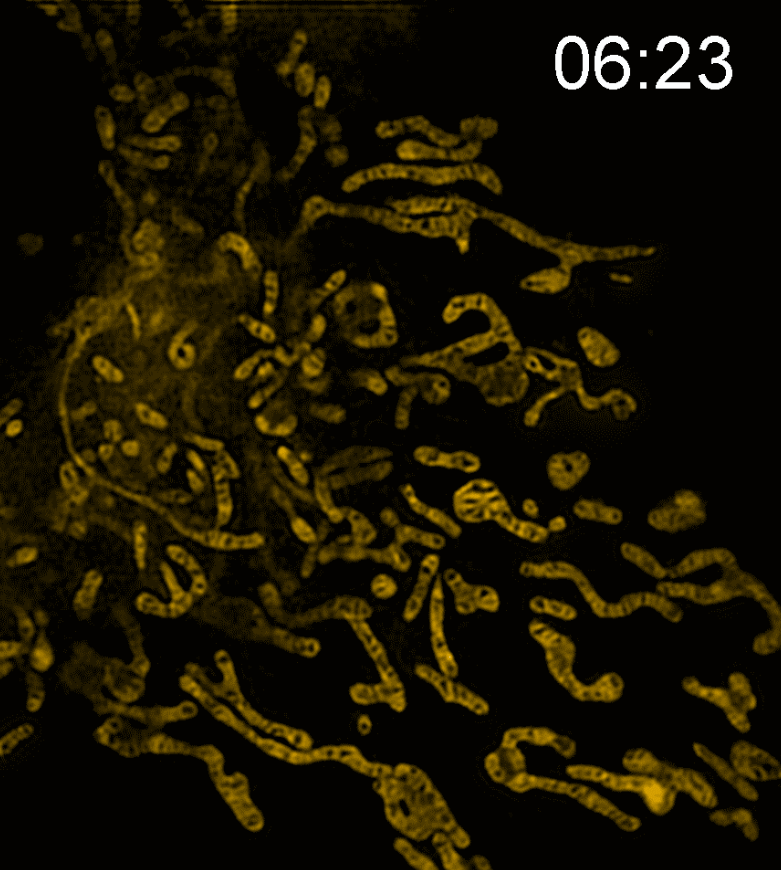
Long-term tracking Based on its low-phototoxicity imaging and full-time tracking strategy, Polar-SIM can perform super-resolution dynamic imaging observation of Live cell mitochondria (light-sensitive samples) for up to tens of minutes
The fast and intelligent large field-of-view image stitching algorithm independently developed by Airy Technologies can achieve seamless splicing in the smallest overlapping areas, reducing photobleaching and phototoxicity.
It can realize different imaging modes, compatible with different magnification objectives and large-field image stitching of different samples.
Airy's unique structured illumination confocal mode enables fast imaging with 150 nm resolution and 50 μm depth in only 2 raw images, making it suitable for the observation of thick tissue samples such as zebrafish, fruit flies, and tissue sections.
Polar-SIM has been developed and optimized for reconstruction algorithms for different modalities, taking into account the fidelity and resolution, while preserving the original information of the sample as much as possible, aiming to provide users with clear, realistic super-resolution images.
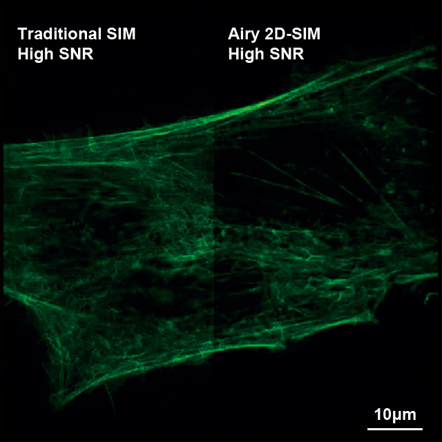
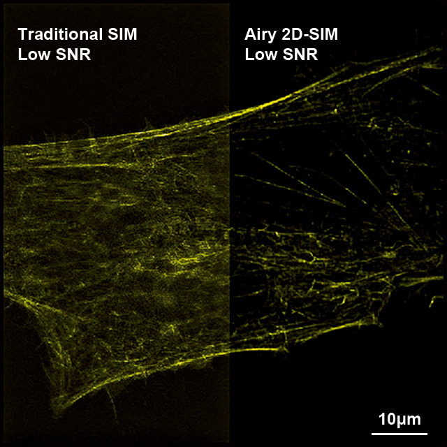
At high signal-to-noise ratio, Airy 2D-SIM behaves similarly to traditional SIMs, reflecting the super-resolution effect of the SIM system. Under low signal-to-noise ratio, Airy 2D-SIM demonstrates better noise robustness through adaptive noise reduction, which can reconstruct samples with extremely low signal-to-noise ratio, maximize the suppression of noise artifacts, and restore the true structure of the sample
Polar-SIM's proprietary algorithm significantly reduces the photon flux required to achieve high-fidelity reconstruction. When the average number of photons exceeds 200, the high-fidelity reconstruction results can be stably output, which greatly reduces the risk of photobleaching and photodamage while ensuring image quality.
AiryTech's exclusive DARK algorithm, designed to overcome strong background interference. It removes background fluorescence from widefield and SIM images, significantly improving the signal-to-noise ratio of images. After processing, widefield images can be rendered with clear optical sectioning effects comparable to confocal for artifact-free, high-resolution imaging in complex areas with strong background signals, such as around the nucleus.
More importantly, DARK provides excellent image quality while significantly reducing the light intensity required for live cell imaging, effectively reducing phototoxic damage and escorting long-term live-cell observation.
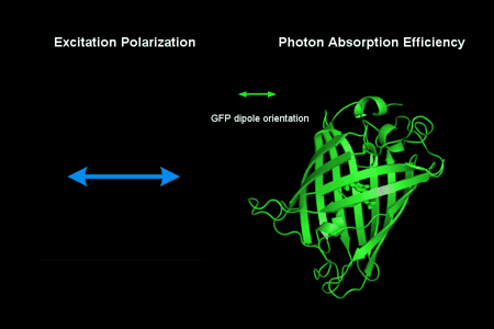
We live in a multi-dimensional world, and fluorescence imaging is no exception. Traditional fluorescence microscopy can only capture fluorophore intensity, but it loses the critical directional dimension. Polar-SIM breaks through this limitation by simultaneously obtaining spatial super-resolution images and fluorescence polarization information, revealing the directional characteristics of the sample at the molecular level: 1. Detection of molecular directionality: Polarization modulation technology is used to accurately analyze the spatial orientation (dipole moment) of fluorescent molecules absorbing/emitting light. 2. Mapping nanoscale ordered structures: Visually display the spatial arrangement patterns and directionality of fluorescent molecules in key structures within cells (such as cytoskeleton, membrane protein complexes). 3. Analyze spatiotemporal dynamic organization: Combine super-resolution (as high as 60 nm) and time-resolution capabilities to clearly track organelle evolution and reveal its highly ordered organization and functional activities in specific directions in the living state (e.g., spindle microtubule polarity, actin directional arrangement). Polar-SIM's polarization dimension, which transcends the limitations of intensity imaging, provides a unique and powerful tool for understanding the regulatory mechanisms of the directional arrangement of molecules in biological activities.


 Public Security Record Number:京公网安备11010802046288号