Sample Source: Airy Technologies
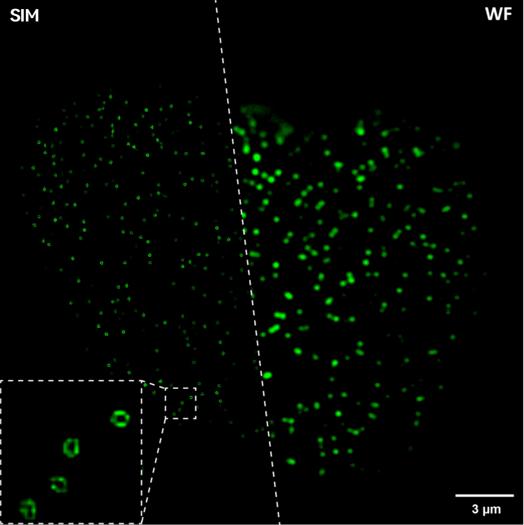
Polar-SIM clearly presents the 60 nm nuclear pore complex structure
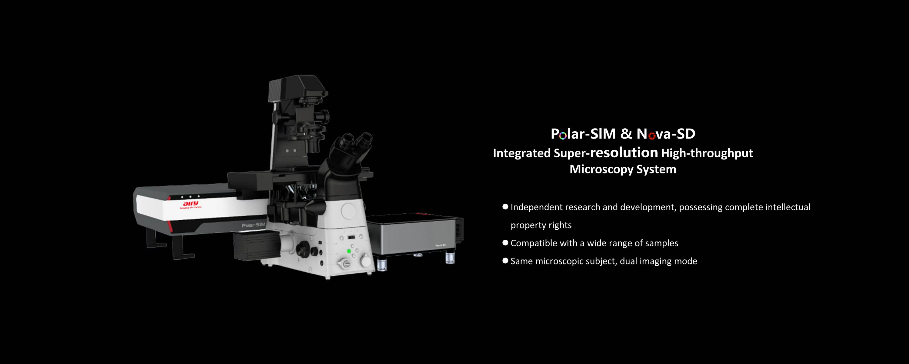
Airy Technologies's fully independently developed integrated super-resolution high-throughput microscopic imaging system innovatively combines SIM super-resolution and Nova-SD spinning disc confocal on a single platform, significantly improving equipment utilization and sharing efficiency: compact design saves space; seamless switching between dual-mode imaging at the same position without sample movement; wide compatibility with various samples to meet the needs of multiple research groups; a single device achieves both super-resolution and deep high-definition imaging, effectively reducing procurement and maintenance costs.
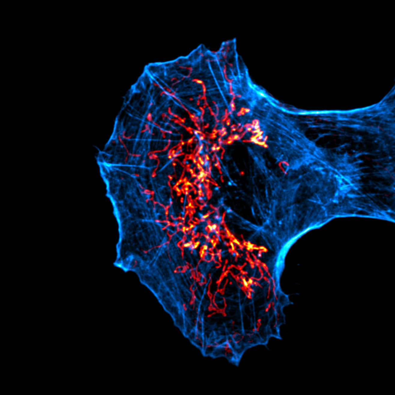
 TIRF-SIM
Total internal reflection structured illumination super-resolution imaging mode
TIRF-SIM
Total internal reflection structured illumination super-resolution imaging mode
TIRF's "zero-background" thin-layer imaging combined with SIM's "non-destructive resolution doubling" provides the ultimate solution for high-fidelity, low-damage, and fast-response nanoscale dynamic studies of living cells, particularly suitable for studying light-sensitive or high-speed dynamic biological processes near the cell membrane.
Data source: Prof. Li Yu Group, Tsinghua University
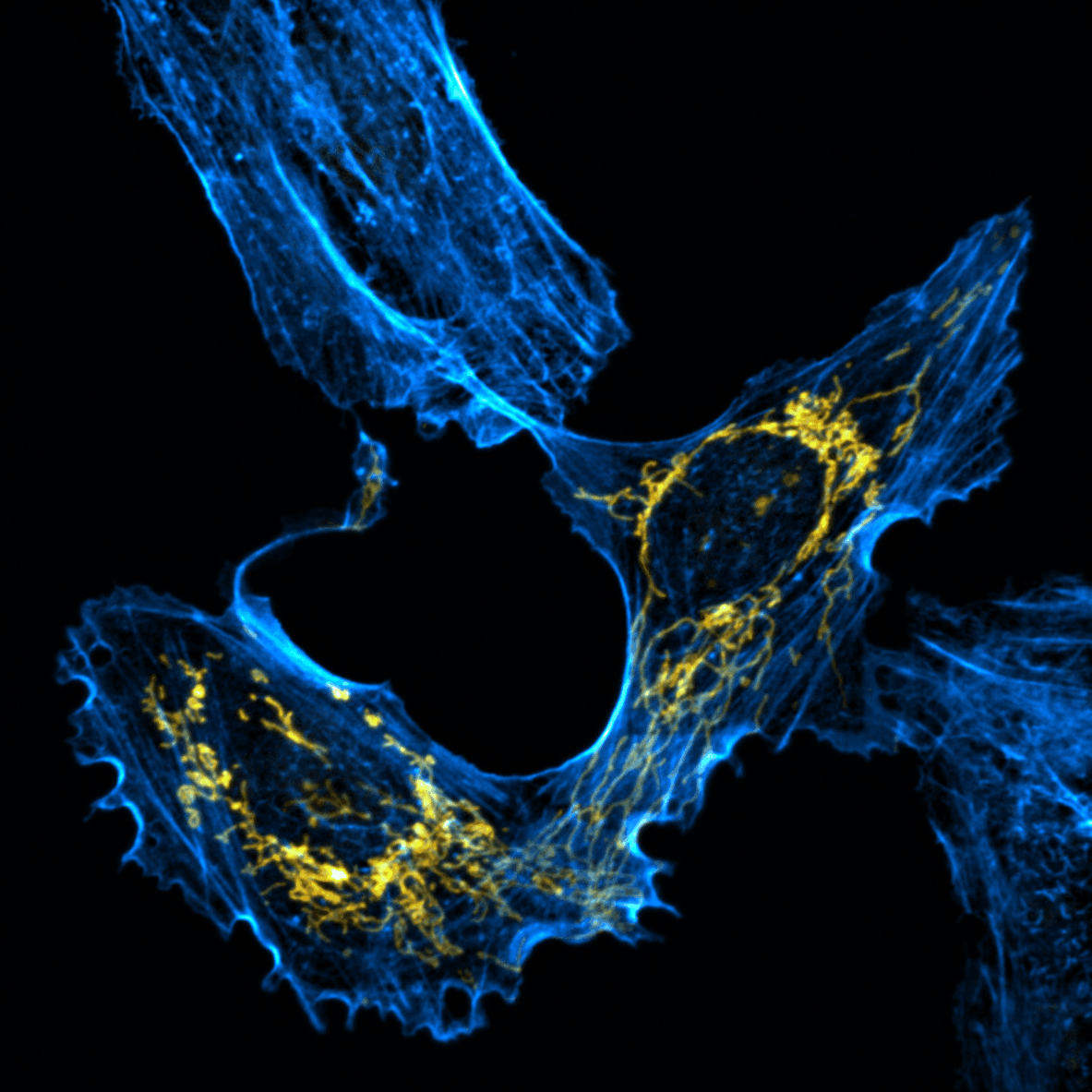
 2D-SIM
Two-dimensional structured illumination super-resolution imaging mode
2D-SIM
Two-dimensional structured illumination super-resolution imaging mode
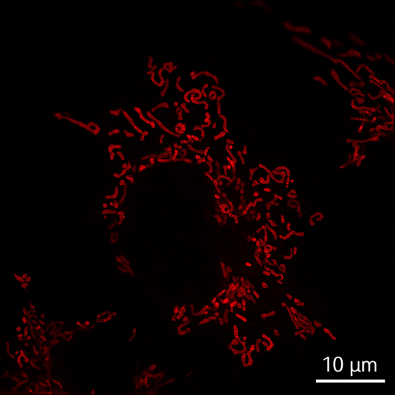
2D-SIM 2D-SIM can clearly resolve subcellular structures without special fluorescent labeling: its low phototoxicity supports second-level dynamic long-term imaging of living cells, avoiding photodamage.
Data source: Hebei University, Gao Baoxiang Group
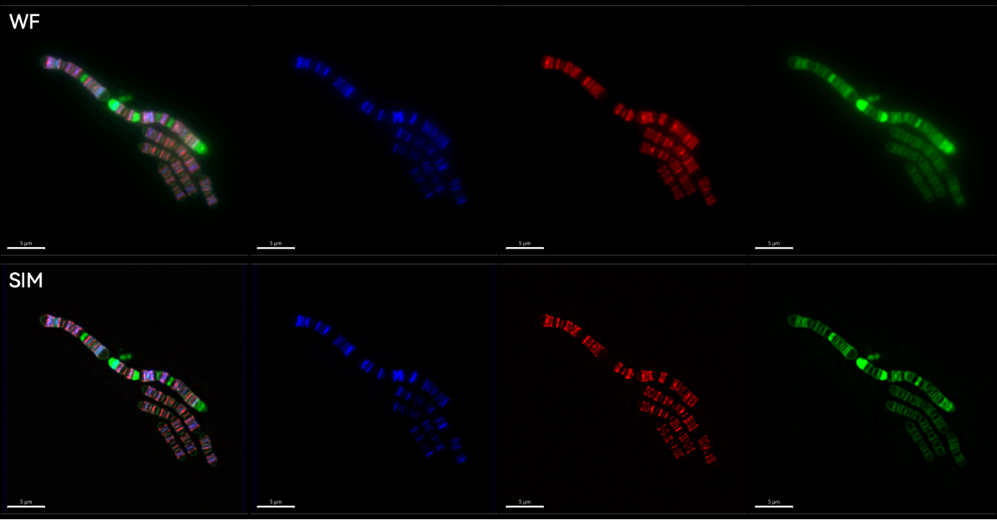
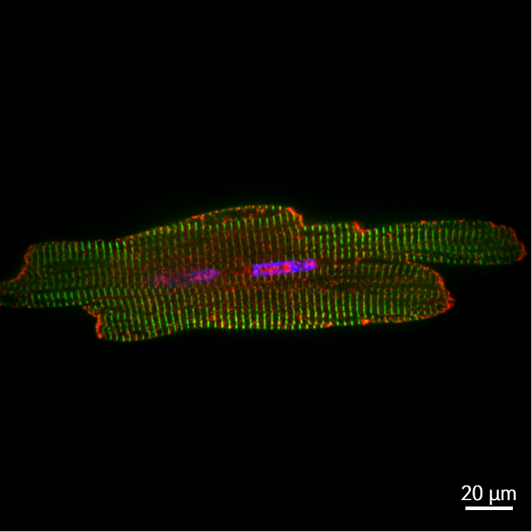
 3D-SIM
3D Structured illumination super-resolution imaging mode
3D-SIM
3D Structured illumination super-resolution imaging mode
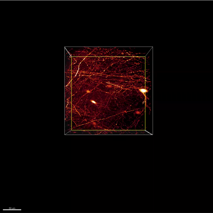
Break through the traditional confocal Z-axis resolution limit, double the 3D imaging accuracy (Z-axis resolution can reach 200 nm), and clearly reconstruct the 3D structure of subcellular organelles such as centrosomes.
Data Source: Airy Technologies
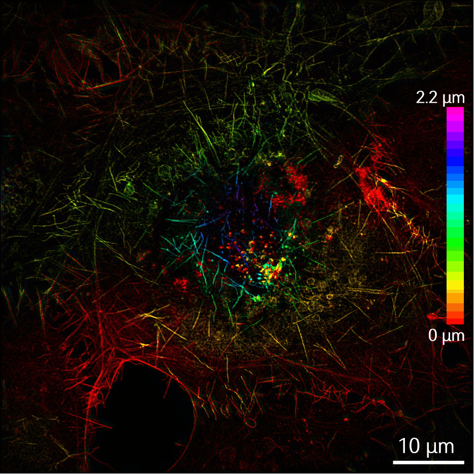
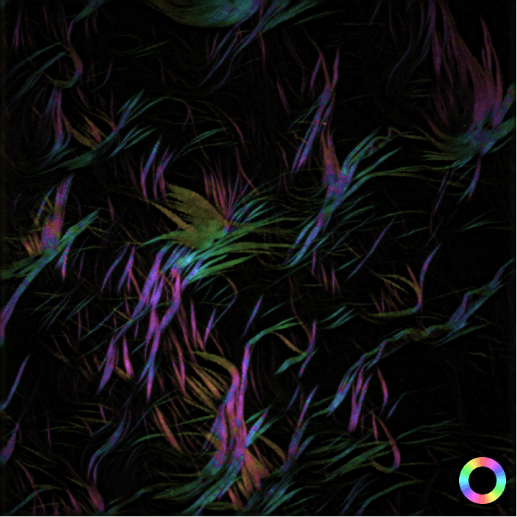 Polar-SIM
Polarized structured illumination super-resolution imaging mode
Polar-SIM
Polarized structured illumination super-resolution imaging mode
Polar-SIM can simultaneously acquire spatial super-resolution images and fluorescence polarization information, revealing the directional characteristics of molecules at the sample level, such as the spatial arrangement patterns and orientation of fluorescent molecules in cytoskeletons, membrane protein complexes, and supramolecular materials.
Data source: Dr. Hailin Fu 's group at Westlake University
 CF1
Spinning disc confocal mode (Higher fluorescent signals)
CF1
Spinning disc confocal mode (Higher fluorescent signals)
The smaller pinhole spacing allows for better collection of fluorescence signals from weak samples, resulting in improved image quality.
Data Source: Airy Technologies
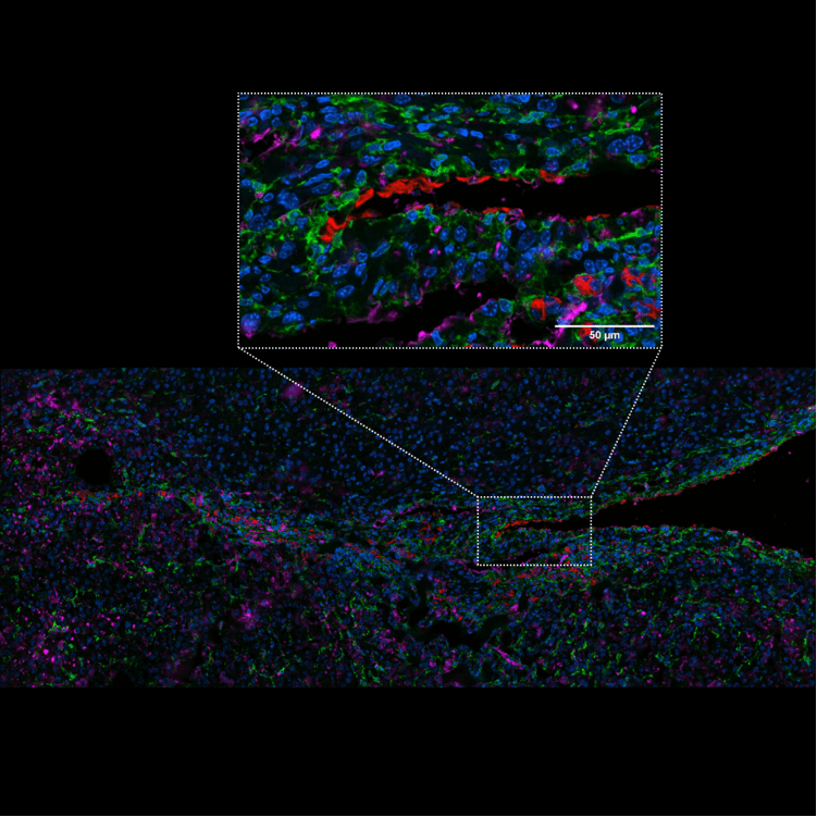
 CF2
Spinning disc confocal mode (better background suppression)
CF2
Spinning disc confocal mode (better background suppression)
Larger pinhole spacing effectively suppresses background signals from thick samples.
Data source: Prof. Ling Tao Group, Xijing Hospital
Based on the user-specified sample imaging area, ensure that the search for the imaging field of view does not exceed the area boundaries, preventing displacement of existing imaging points.
Application scenario: In multi-point long-term observations, if the field of view is moved outside the bottom chamber of the confocal petri dish, confirmed points may shift.
Preview ultra-wide field imaging based on user-specified areas, to quickly confirm subsequent imaging points and areas through preview images, and reduce field-of-view searching time.
Application scenario: Point-by-point scanning of sample areas to find imaging fields with co-expressed dual/multi-target proteins is time-consuming and inefficient.
When the required imaging area is larger than the optical field of view, seamless stitching is supported to generate large-format images.
Application scenarios: panoramic imaging of small intestine, liver, zebrafish, etc.
Automatically plan the optimal collection path based on multiple user-specified points to achieve efficient coverage.
Application scenarios: Multi-point long-term observation or imaging of the same cells before and after drug administration.
Users can specify multiple imaging points for four-dimensional imaging in xyzt coordinates.
Application scenarios: Multi-point long-term observation, where the observed object may undergo vertical displacement along the Z-axis.
For the same imaging area, both confocal imaging and SIM super-resolution imaging modes are supported.
Application scenario: In confocal imaging, two proteins are found to co-localize, but limited by the resolution of confocal imaging, the SIM mode can be used to further clarify the details of co-localization.
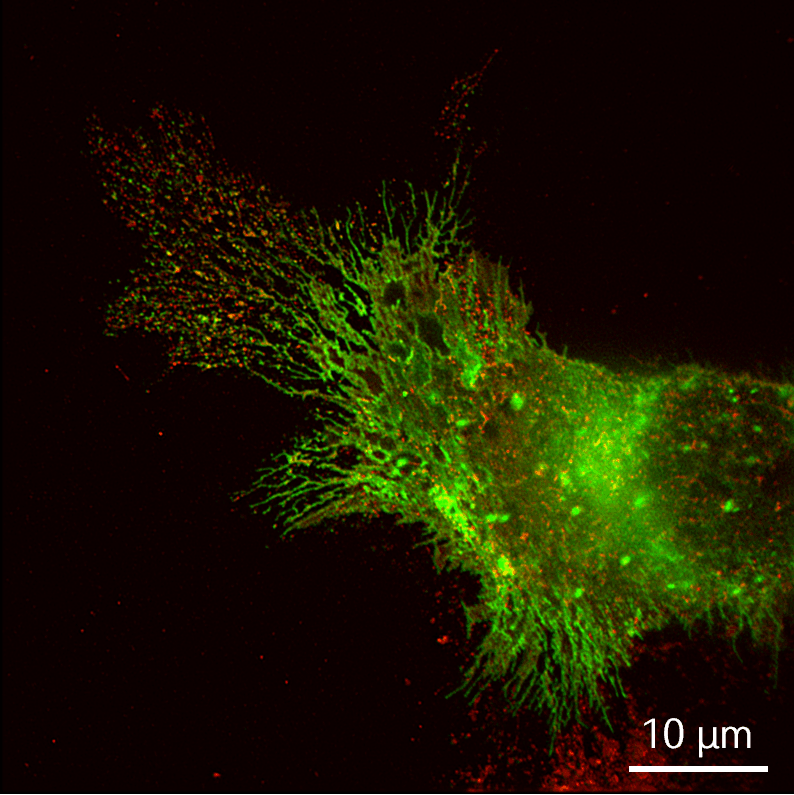
Airy Technologies provides multi-color rapid simultaneous imaging solutions, helping users perform long-term observations of multiple subcellular structures in motion within living cells, truly reflecting the interaction processes of organelles.
Application scenario: Observing the involvement of connexins in dynamic processes of the endoplasmic reticulum.
Fluorescence anisotropy measurement imaging technology helps users analyze molecular interactions, protein conformational changes, and monitor cell membrane states.
Application scenario: Observe the changes in orientation of cytoskeletal proteins before and after drug administration.


 Public Security Record Number:京公网安备11010802046288号